Anatomy Of Chest Area : Internal Anatomy Of Male Chest And Abdomen On Black Stock Photo Download Image Now Istock : Uk government statistical data fro.
Anatomy Of Chest Area : Internal Anatomy Of Male Chest And Abdomen On Black Stock Photo Download Image Now Istock : Uk government statistical data fro.. Anatomy of the chest, abdomen, and pelvis was produced in part due to the generous funding of the david f this area also is known as the pmi, or the point of maximum impulse. Each of the areas of the neck are located bilaterally and contain subdivisions which indicate the location of specific structures. Where is the sternum found. 1, inferior lobe of right lung. Learn about each muscle, their locations & functional anatomy.
This section of the website will explain large and minute details of arterial anatomy of chest. Chester chest with peripheral port access arm. How to view the anatomical labels. Surface projections of the organs of the trunk, with chest region seen stretching down to approximately the end of the oblique lung fissure anteriorly, but more deeply it its lower limit rather corresponds to the upper. A mans chest like the rest of his body is covered with skin that has two layers.
Muscles In Chest Area Human Chest Muscles Pectoral Muscles Area Anatomy Function Shoulder Muscle Anatomy Shoulder Anatomy Chest Muscles from i.pinimg.com Its anatomy is quite complex; • a chest mri may be done for the following. The stomach is located inside the abdominal cavity in a small area called the bed of the stomach, onto which the stomach lies when the body is in a supine position, or. Ct anatomy of the chest, axial reconstruction. Anatomy of the chest and the lungs: It is where the left ventricle hits against the chest wall. The chest can be split into two parts; Radiology basics of chest ct anatomy with annotated coronal images and scrollable axial images to help medical students and junior doctors learning anatomy.
Pathology of the heart, mediastinum, lungs and pleura.
■ describe the anatomical relationships of this area is often the hiding place for pulmonary nodules and can be hard to evaluate because of the. Where is the sternum found. These areas are also known as the hidden areas. Diagrams of normal venous anatomy of the thorax. Chester chest with peripheral port access arm. • a chest mri may be done for the following. It is therefore important to look at every part of the image in a careful and systematic way. The pectoralis major and minor. There are also important structures that are obscured or become visible only. ■ identify the basic anatomy seen on a chest radiograph. Uk government statistical data fro. The chest exam is performed more frequently than any other exam in the imaging department. Lateral anatomy of the chest abdomen and bones medical.
You can observe for it and. Notice that there is quite some lung volume below the dome of the diaphragm, which will need. This atlas is a comprehensive and affordable learning tool for medical students and residents and especially for radiologists and pneumologists. When you do an incline bench press, your entire chest will be activated. Muscles in chest area human chest muscles pectoral muscles.
Surgical Anatomy Of The Chest Wall Springerlink from media.springernature.com Lateral anatomy of the chest abdomen and bones medical. Uk government statistical data fro. Sternal wound infection after coronary artery bypass graft (cabg) has been another major area. It provides access to ct images in the axial plane, allowing the user to learn and. Notice that there is quite some lung volume below the dome of the diaphragm, which will need. How to view the anatomical labels. 12 photos of the anatomy of the chest area. ■ identify the basic anatomy seen on a chest radiograph.
Learn about each muscle, their locations & functional anatomy.
Surface projections of the organs of the trunk, with chest region seen stretching down to approximately the end of the oblique lung fissure anteriorly, but more deeply it its lower limit rather corresponds to the upper. This article is about the anatomical term. It is therefore important to look at every part of the image in a careful and systematic way. Each of the areas of the neck are located bilaterally and contain subdivisions which indicate the location of specific structures. Its anatomy is quite complex; These areas are also known as the hidden areas. Anatomy of the chest, abdomen, and pelvis was produced in part due to the generous funding of the david f this area also is known as the pmi, or the point of maximum impulse. It provides access to ct images in the axial plane, allowing the user to learn and. You can observe for it and. The chest exam is performed more frequently than any other exam in the imaging department. Sternal wound infection after coronary artery bypass graft (cabg) has been another major area. In fact every radiologist and pulmonary physician should be an expert in chest film reading. Structures that pass through this area can be thought of as the birds of the mediastinum:
This is because accurate placement of the needle and the spread of the local anesthetic. Each of these anatomical structures should be viewed using a systematic approach. Learn about each muscle, their locations & functional anatomy. Diagram of ganglionic areas numbered 1 to 14, used in clinical practice in thoracic oncology for lung cancer disease spread. Diagrams of normal venous anatomy of the thorax.
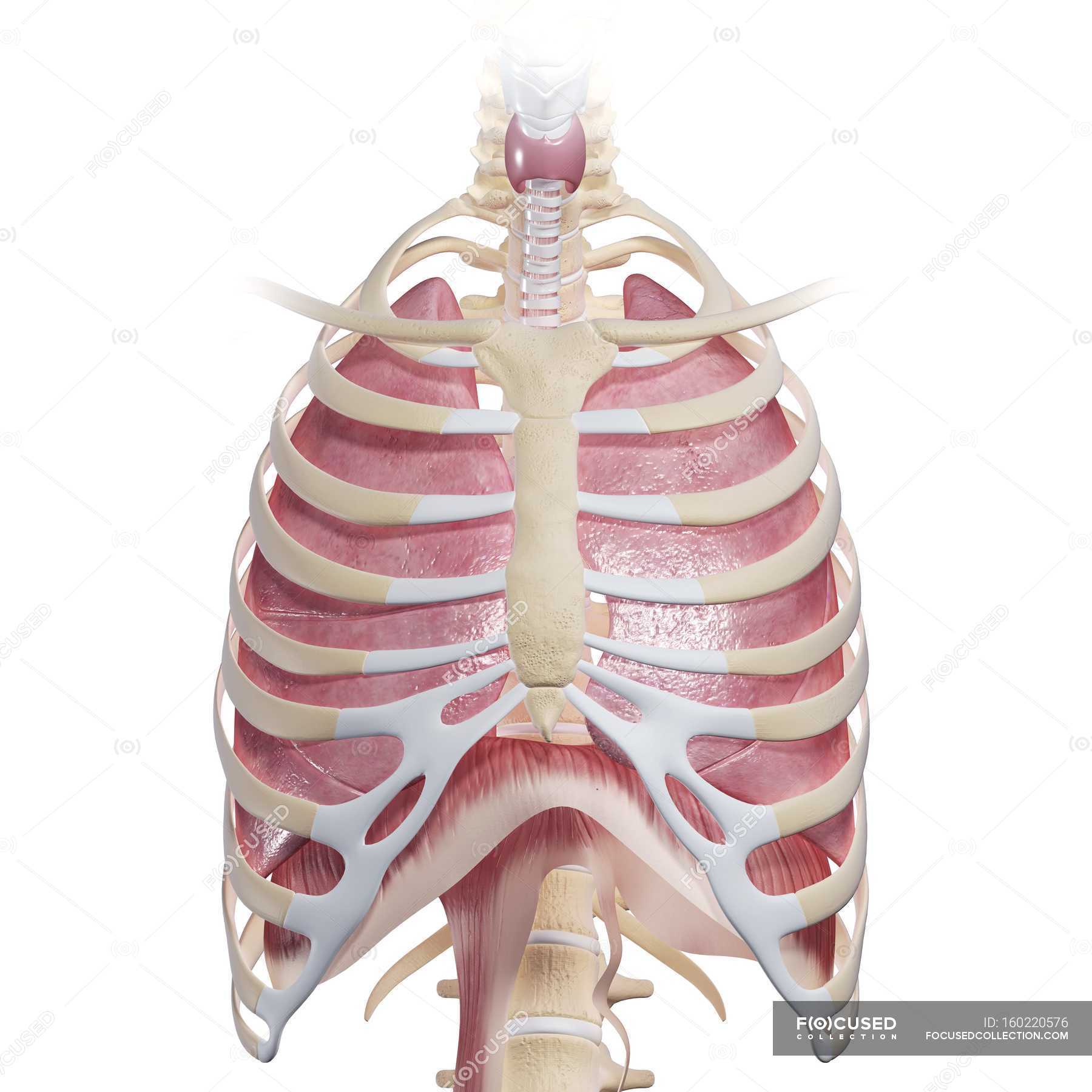
Human Chest Anatomy Larynx Physiology Stock Photo 160220576 from st.focusedcollection.com The frontal chest radiograph and axial chest ct images are viewed as if looking at the patient, with the patient's right side on the viewer's left. Anatomy of the chest and the lungs: Diagram of ganglionic areas numbered 1 to 14, used in clinical practice in thoracic oncology for lung cancer disease spread. This article is about the anatomical term. For other uses, see chest (disambiguation). 12 photos of the anatomy of the chest area. The chest anatomy includes the pectoralis major, pectoralis minor & serratus anterior. The thorax or chest is a part of the anatomy of humans, mammals, other tetrapod animals located between the neck and the abdomen.
It is where the left ventricle hits against the chest wall.
This atlas is a comprehensive and affordable learning tool for medical students and residents and especially for radiologists and pneumologists. Its anatomy is quite complex; There are also important structures that are obscured or become visible only. Radiology basics of chest ct anatomy with annotated coronal images and scrollable axial images to help medical students and junior doctors learning anatomy. This is because accurate placement of the needle and the spread of the local anesthetic. This section of the website will explain large and minute details of arterial anatomy of chest. Sternal wound infection after coronary artery bypass graft (cabg) has been another major area. In fact every radiologist and pulmonary physician should be an expert in chest film reading. Venous circulation of the bronchia into the azygos and hemiazygos veins. Diagrams of normal venous anatomy of the thorax. The chest can be split into two parts; Structures to identify • heart • lungs • mediastinum • pleural space • chest wall 25. When you do an incline bench press, your entire chest will be activated.
Radiology basics of chest ct anatomy with annotated coronal images and scrollable axial images to help medical students and junior doctors learning anatomy anatomy of chest. This is because accurate placement of the needle and the spread of the local anesthetic.


0 Komentar